Oligodendroglioma is a type of diffusely infiltrating glioma, most common in adults between 25 and 45 years old. Neuroimaging constitutes an essential component of both diagnostic evaluation and surgical planning. While conventional imaging modalities effectively delineate tumor location and extent, they possess inherent limitations in detecting early pathological changes. Ultrasound imaging enjoys widespread clinical adoption owing to its excellent tissue penetration, lack of ionizing radiation, real-time imaging capacity, and equipment portability. Intraoperative ultrasound tools like contrast-enhanced ultrasound (CEUS) have become crucial for real-time tumor mapping. CEUS uses microbubble contrast agents to enhance tumor visualization by highlighting perfusion differences, improving boundary definition and residual tumor detection. Super-resolution ultrasound (SR-US) has emerged as an innovative imaging modality capable of high-resolution vascular mapping and functional assessment. Given these capabilities, this article presents a CEUS-guided surgical case, demonstrating its role in optimizing tumor resection while preserving brain function. A 33-year-old female patient was admitted to the hospital with a three-day history of an intracranial mass detected during a routine physical examination. MRI revealed a cysto-solid mass in the right frontal lobe, suggesting oligodendroglioma. The patient underwent a right frontal lobe resection, during which intraoperative ultrasound, CEUS, and SR-US imaging were performed. These imaging techniques revealed a cysto-solid mass with no blood vessels within the tumor and a rich microvascular structure in the surrounding brain tissue. The tumor was resected, and pathological examination confirmed the diagnosis of oligodendroglioma (WHO Grade II). MRI is the preferred diagnostic method, while CEUS and SR-US imaging have significant value in the diagnosis and treatment of brain tumors, improving diagnostic accuracy and surgical resection rates.
| Published in | Journal of Surgery (Volume 13, Issue 5) |
| DOI | 10.11648/j.js.20251305.11 |
| Page(s) | 120-125 |
| Creative Commons |
This is an Open Access article, distributed under the terms of the Creative Commons Attribution 4.0 International License (http://creativecommons.org/licenses/by/4.0/), which permits unrestricted use, distribution and reproduction in any medium or format, provided the original work is properly cited. |
| Copyright |
Copyright © The Author(s), 2025. Published by Science Publishing Group |
Oligodendroglioma, Ultrasound, Microbubbles, Contrast, Super-Resolution Ultrasound Imaging
| [1] | Tork CA, Hall WA, Atkinson C. Oligodendroglioma. 2025 Apr 3. In: StatPearls [Internet]. Treasure Island (FL): StatPearls Publishing; 2025 Jan-. PMID: 32644610. |
| [2] | Louis DN, Ohgaki H, Wiestler OD, et al. The 2007 WHO classification of tumours of the central nervous system. Acta Neuropathol 2007; 114(2): 97-109. |
| [3] | Zulfiqar M, Dumrongpisutikul N, Intrapiromkul J, et al. Detection of intratumoral calcification in oligodendrogliomas by susceptibility-weighted MR imaging. AJNR Am J Neuroradiol 2012; 33: 858-64. |
| [4] | Chourmouzi, D., Papadopoulou, E., Marias, K., & Drevelegas, A. (2014). Imaging of brain tumors. Surgical oncology clinics of North America, 23(4), 629-684. |
| [5] | Lee EJ, Lee SK, Agid R, et al. Preoperative grading of presumptive low-grade astrocytomas on MR imaging: diagnostic value of minimum apparent diffusion coefficient. AJNR Am J Neuroradiol 2008; 29: 1872-7. |
| [6] | V. Costalat, T. G. Jovin, J. F. Albucher, C. Cognard, H. Henon, N. Nouri, et al., Trial of thrombectomy for stroke with a large infarct of unrestricted size, N. Engl. J. Med. 390 (18) (2024) 1677-1689, |
| [7] | G. Broocks, A. Kemmling, S. Teßarek, R. McDonough, L. Meyer, T. D. Faizy, et al., Quantitative lesion water uptake as stroke imaging biomarker: a tool for treatment selection in the extended time window? Stroke 53 (1) (2022) 201-209, |
| [8] | M. Zhang, Z. Wang, C. Wang, Y. Wu, Z. Li, Z. Liu, Visualizing oxidative stress level for timely assessment of ischemic stroke via a ratiometric near-infrared-II luminescent nanoprobe, ACS Nano 15 (7) (2021) 11940-11952, |
| [9] | C. Song, L. Liao, Y. Bin, Z. He, J. Hua, S. Zhao, et al., Visualization diagnosis of acute cerebral ischemia via sulfane sulfur-activated photoacoustic imaging, Chem. Commun. 60 (9) (2024) 1112-1115, |
| [10] | P. Liu, S. Lan, D. Gao, D. Hu, Z. Chen, Z. Li, et al., Targeted blood-brain barrier penetration and precise imaging of infiltrative glioblastoma margins using hybrid cell membrane-coated ICG liposomes, J. Nanobiotechnol. 22 (1) (2024) 603, |
| [11] | J. Xu, N. Zhu, Y. Du, T. Han, X. Zheng, J. Li, et al., Biomimetic NIR-II fluorescent proteins created from chemogenic protein-seeking dyes for multicolor deep-tissue bioimaging, Nat. Commun. 15 (1) (2024) 2845, |
| [12] | Z. Zhang, Y. Du, X. Shi, K. Wang, Q. Qu, Q. Liang, et al., NIR-II light in clinical oncology: opportunities and challenges, Nat. Rev. Clin. Oncol. 21 (6) (2024) 449-467, |
| [13] | W. Pan, M. Rafiq, W. Haider, Y. Guo, H. Wang, M. Xu, et al., Recent advances in NIR-II fluorescence/photoacoustic dual-modality imaging probes, Coord. Chem. Rev. 514 (2024) 215907, |
| [14] | L. Yuan, Q. Li, Z. Li, Effects of ultrasound for bio-applications, Advanced Sensor Research 3 (8) (2024) 2300199, |
| [15] | X. Liu, J. Wang, Y. Wu, M. Wu, J. Song, Ultrasound activated probe for disease imaging and therapy In-Vivo, Adv. Drug Deliv. Rev. 205 (2024) 115158, |
| [16] | J. Yan, B. Huang, J. Tonko, M. Toulemonde, J. Hansen-Shearer, Q. Tan, et al., Transthoracic ultrasound localization microscopy of myocardial vasculature in patients, Nat. Biomed. Eng. 8 (6) (2024) 689-700, |
| [17] | K. Christensen-Jeffries, O. Couture, P. A. Dayton, Y. C. Eldar, K. Hynynen, F. Kiessling, et al., Super-resolution ultrasound imaging, Ultrasound Med. Biol. 46 (4) (2020) 865-891, |
| [18] | J. Arendt Jensen, M. Amin Naji, S. Kazmarek PraeSius, I. Taghavi, M. Schou, L. Naur Hansen, et al., Super-resolution ultrasound imaging using the erythrocytes- Part I: density images, IEEE Trans. Ultrason. Ferroelectrics Freq. Control 71 (8) (2024) 925-944, |
| [19] | M. A. Naji, I. Taghavi, M. Schou, S. K. Praesius, L. N. Hansen, N. S. Panduro, et al., Super-resolution ultrasound imaging using the erythrocytes-Part II: velocity images, IEEE Trans. Ultrason. Ferroelectrics Freq. Control 71 (8) (2024) 945-959, |
| [20] | Tork CA, Atkinson C. Oligodendroglioma. In: StatPearls. Treasure Island (FL): StatPearls Publishing; August 28, 2023. |
| [21] | Joyner DA, Garrett J, Batchala PP, et al. MRI features predict tumor grade in isocitrate dehydrogenase (IDH)-mutant astrocytoma and oligodendroglioma. Neuroradiology. 2023; 65(1):121-129. |
| [22] | Smits M. Imaging of oligodendroglioma. Br J Radiol. 2016; 89(1060):20150857. |
| [23] | Kruger K, Silwal-Pandit L, Wik E, Straume O, Stefansson IM, Borgen E, Garred Ø, Naume B, Engebraaten O, Akslen LA. Baseline microvessel den sity predicts response to neoadjuvant bevacizumab treatment of locally advanced breast cancer. Sci Rep. 2021; 11: 3388. |
| [24] | Maxeiner A, Fischer T, Schwabe J, Baur ADJ, Stephan C, Peters R, Slowinski T, von Laffert M, Marticorena Garcia SR, Hamm B, Jung EM. Contrast enhanced US (CEUS) and quantitative perfusion analysis in patients with suspicion for prostate cancer. Ultraschall Med. 2019; 40: 340-8. |
| [25] | Yunkai Z, Yaqing C, Jun J, Tingyue Q, Weiyong L, Yuehong Q, Wenbin G, Lifeng W, Jun Q. Comparison of contrast-enhanced US targeted biopsy versus standard systematic biopsy for clinically significant prostate cancer detection: results of a prospective cohort study with 1024 patients. World J Urol. 2019; 37(5): 805-11. |
| [26] | Yan L, Bai C, Zheng Y, et al. Study on the Application of Super-Resolution Ultrasound for Cerebral Vessel Imaging in Rhesus Monkeys. Front Neurol. 2021; 12: 720320. Published 2021 Nov 17. |
| [27] | Xia S, Zheng Y, Hua Q, et al. Super-resolution ultrasound and microvasculomics: a consensus statement. Eur Radiol. 2024; 34(11):7503-7513. |
| [28] | Chavignon, A., Hingot, V., Orset, C., Vivien, D., & Couture, O. (2022). 3D transcranial ultrasound localization microscopy for discrimination between ischemic and hemorrhagic stroke in early phase. Scientific reports, 12(1), 14607. |
| [29] | Schwarz, S., Denis, L., Nedoschill, E., Buehler, A., Danko, V., Hilger, A. C., Brevis Nuñez, F., Dürr, N. R., Schlunz-Hendann, M., Brassel, F., Felderhoff-Müser, U., Reutter, H., Woelfle, J., Jüngert, J., Dohna-Schwake, C., Bruns, N., Regensburger, A. P., Couture, O., Mandelbaum, H., & Knieling, F. (2025). Ultrasound Super-Resolution Imaging of Neonatal Cerebral Vascular Reorganization. Advanced science (Weinheim, Baden-Wurttemberg, Germany), 12(12), e2415235. |
APA Style
Liu, X., Song, X., Liu, C., Zheng, Y. (2025). Super-Resolution Ultrasound Imaging of Oligodendroglioma: A Case Report and Literature Review. Journal of Surgery, 13(5), 120-125. https://doi.org/10.11648/j.js.20251305.11
ACS Style
Liu, X.; Song, X.; Liu, C.; Zheng, Y. Super-Resolution Ultrasound Imaging of Oligodendroglioma: A Case Report and Literature Review. J. Surg. 2025, 13(5), 120-125. doi: 10.11648/j.js.20251305.11
@article{10.11648/j.js.20251305.11,
  author = {Xinyu Liu and Xue Song and Cun Liu and Yanling Zheng},
  title = {Super-Resolution Ultrasound Imaging of Oligodendroglioma: A Case Report and Literature Review
},
  journal = {Journal of Surgery},
  volume = {13},
  number = {5},
  pages = {120-125},
  doi = {10.11648/j.js.20251305.11},
  url = {https://doi.org/10.11648/j.js.20251305.11},
  eprint = {https://article.sciencepublishinggroup.com/pdf/10.11648.j.js.20251305.11},
  abstract = {Oligodendroglioma is a type of diffusely infiltrating glioma, most common in adults between 25 and 45 years old. Neuroimaging constitutes an essential component of both diagnostic evaluation and surgical planning. While conventional imaging modalities effectively delineate tumor location and extent, they possess inherent limitations in detecting early pathological changes. Ultrasound imaging enjoys widespread clinical adoption owing to its excellent tissue penetration, lack of ionizing radiation, real-time imaging capacity, and equipment portability. Intraoperative ultrasound tools like contrast-enhanced ultrasound (CEUS) have become crucial for real-time tumor mapping. CEUS uses microbubble contrast agents to enhance tumor visualization by highlighting perfusion differences, improving boundary definition and residual tumor detection. Super-resolution ultrasound (SR-US) has emerged as an innovative imaging modality capable of high-resolution vascular mapping and functional assessment. Given these capabilities, this article presents a CEUS-guided surgical case, demonstrating its role in optimizing tumor resection while preserving brain function. A 33-year-old female patient was admitted to the hospital with a three-day history of an intracranial mass detected during a routine physical examination. MRI revealed a cysto-solid mass in the right frontal lobe, suggesting oligodendroglioma. The patient underwent a right frontal lobe resection, during which intraoperative ultrasound, CEUS, and SR-US imaging were performed. These imaging techniques revealed a cysto-solid mass with no blood vessels within the tumor and a rich microvascular structure in the surrounding brain tissue. The tumor was resected, and pathological examination confirmed the diagnosis of oligodendroglioma (WHO Grade II). MRI is the preferred diagnostic method, while CEUS and SR-US imaging have significant value in the diagnosis and treatment of brain tumors, improving diagnostic accuracy and surgical resection rates.
},
 year = {2025}
}
TY  - JOUR
T1  - Super-Resolution Ultrasound Imaging of Oligodendroglioma: A Case Report and Literature Review

AU  - Xinyu Liu
AU  - Xue Song
AU  - Cun Liu
AU  - Yanling Zheng
Y1  - 2025/09/05
PY  - 2025
N1  - https://doi.org/10.11648/j.js.20251305.11
DO  - 10.11648/j.js.20251305.11
T2  - Journal of Surgery
JF  - Journal of Surgery
JO  - Journal of Surgery
SP  - 120
EP  - 125
PB  - Science Publishing Group
SN  - 2330-0930
UR  - https://doi.org/10.11648/j.js.20251305.11
AB  - Oligodendroglioma is a type of diffusely infiltrating glioma, most common in adults between 25 and 45 years old. Neuroimaging constitutes an essential component of both diagnostic evaluation and surgical planning. While conventional imaging modalities effectively delineate tumor location and extent, they possess inherent limitations in detecting early pathological changes. Ultrasound imaging enjoys widespread clinical adoption owing to its excellent tissue penetration, lack of ionizing radiation, real-time imaging capacity, and equipment portability. Intraoperative ultrasound tools like contrast-enhanced ultrasound (CEUS) have become crucial for real-time tumor mapping. CEUS uses microbubble contrast agents to enhance tumor visualization by highlighting perfusion differences, improving boundary definition and residual tumor detection. Super-resolution ultrasound (SR-US) has emerged as an innovative imaging modality capable of high-resolution vascular mapping and functional assessment. Given these capabilities, this article presents a CEUS-guided surgical case, demonstrating its role in optimizing tumor resection while preserving brain function. A 33-year-old female patient was admitted to the hospital with a three-day history of an intracranial mass detected during a routine physical examination. MRI revealed a cysto-solid mass in the right frontal lobe, suggesting oligodendroglioma. The patient underwent a right frontal lobe resection, during which intraoperative ultrasound, CEUS, and SR-US imaging were performed. These imaging techniques revealed a cysto-solid mass with no blood vessels within the tumor and a rich microvascular structure in the surrounding brain tissue. The tumor was resected, and pathological examination confirmed the diagnosis of oligodendroglioma (WHO Grade II). MRI is the preferred diagnostic method, while CEUS and SR-US imaging have significant value in the diagnosis and treatment of brain tumors, improving diagnostic accuracy and surgical resection rates.

VL  - 13
IS  - 5
ER  -
Department of Clinical Medicine, First Clinical Medical College of Zhengzhou University, Zhengzhou, China
Department of Ultrasound, Jinan Central Hospital Affiliated to Shandong First Medical University, Jinan, China
Department of Ultrasound, Jinan Central Hospital Affiliated to Shandong First Medical University, Jinan, China
School of Mathematical Science, University of Jinan, Jinan, China
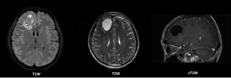
Figure 1. Transverse contrast-enhanced T1-weighted (T1W), T2-weighted (T2W), and contrast-enhanced T1-weighted (cT1W) images of the right frontal lobe oligodendroglioma.
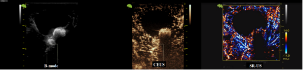
Figure 2. B-mode image showing a cysto-solid mass in the right frontal lobe, predominantly cystic, with a clear boundary and good internal acoustic transparency. A nodular hyperechoic structure with acoustic shadowing is observed on the deep cyst wall. CEUS reveals non-enhancement within the anechoic areas of the tumor, with heterogeneous enhancement observed in the surrounding brain parenchyma. SR-US imaging of the lesion demonstrates a well-circumscribed and dense microvasculature. The color bar represents blood flow direction, with red and blue indicating flow toward and away from the transducer, respectively.
Information

