2. Methodology
This was an experimental, cross-sectional, descriptive and analytical study that took place over two years in Yaoundé (Cameroon). It was approved by the Ethics Committee of the Yaoundé University Hospital in 2015. To which is added the multidisciplinary laboratory of pharmaceutical sciences FMSB of the University of Yaoundé I.
2.1. Material
The material used for this study included:
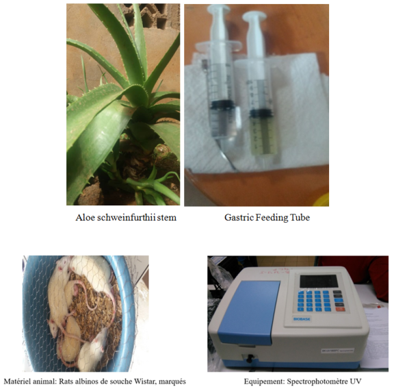
1. Plant material: Aloe schweinfurthii gel identified by the national herbarium of Cameroon
2. Solvents: Aqueous eosin and physiological serum;
3. Human material: Periodontal ligament of teeth extracted atraumatically;
4. Animal material: consisting of male and female albino rats, Rattus norvegicus of the Wistar strain.
5. Laboratory equipment, clinical examination and measuring devices, spectrophotometer and magnetic stirrer. The measuring devices, the spectrophotometer and the magnetic stirrer.
Figure 1. Equipment used during the performance of acute and subacute toxicity acts.
Figure 2. Volumes of test solution (2000 and 5000) mg/kg body weight.
2.2. Methods
2.2.1. Preparation of Volumes of Test Solution
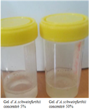
The dilution concentrations with a constant volume of distilled water of 100 milliliters were obtained by calculating the mass ratio of the A. schweinfurthii gel used to the volume of water. Based on the observed conservation results of live cells after 24 hours of more than 87 to 99% on all concentrations in a previous study by Mengong et al
| [11] | Mengong Moneboulou HP, Nokam Abena ME, Ndjoh J, Essama Eno B, Zing S, Bengondo C, Kattie Louka A. Recherche d’un milieu de conservation optimal de la dent permanente immature expulsée par traumatisme. Rev Col Odonto-Stomatolo Afr Chir Maxillo-fac.2020; P 11-17. |
[11]
; A choice of toxicity study was made on the two extremes studied, namely the concentration of 5 and 50% studied. To ensure that, when the preservation device is set up, priority is given to the lowest and least toxic concentration in order to optimize the balance between the benefits of the drug and its risks.
The doses of 2000 mg/kg and 5000 mg/kg were chosen based on standardized guidelines set the OECD (Organisation for Economic Cooperation and Development) gide line. These doses are large enough to reveal potential toxic effects after short-term high-dose exposure or long term. By examining the outcomes, researchers can better understand the safety profile of Aloe schweinfurthii. The higher the dose, the more likely it is to unearth any adverse effects.
D = test dose (mg/kg)
C = concentration of A. S (mg/mL)
P = weight of the animal (kg)
V = volume of solution to be administered (mL)
2.2.2. Evaluation of the Frost Toxicity of A.Schweinfurthii
(i). Evaluation of the Acute Oral Toxicity in Vitro of A.schweinfurthii Gel
The acute oral toxicity in vitro of aloe vera Schweinfurthii gel was conducted according to OECD guideline 425. It consisted in highlighting the maximum tolerated concentration of A.schweinfurthii gel / oral administration in single dose / 24 h. It was conducted on 9 female rats randomly divided into 3 groups aged 6-8 weeks with an average weight of (94 ± 10 g). This study was conducted in albino rats of Wistar strains with therapeutic doses of 5% and 50% having shown a good conservation activity of the immature permanent tooth.
Protocol
The acute oral toxicity study consisted in determining the lethal dose 50 (LD50) and the toxic effects of the aqueous extract of Aloe schweinfurthii Baker administered as a single dose to female rats. The methodology used was that described by OECD 425 (Organisation for Economic Co-operation and Development) with some modifications
. The experiment was conducted on 9 female rats weighing between 94 and 112 g. The animals were randomly selected, marked for identification purposes and randomly distributed into three groups of 3 animals each. The dose to be administered was calculated according to the fasting body weight of each animal according to the formula for calculating the initial volume according to weight. The animals were fasted for 12 hours but were hydrated the night before the experiment. Doses of
Aloe schweinfurthii Baker mucilaginous gel of 2000 and 5000 mg/kg were administered orally as a single dose to rats in groups 2 and 3. Administration was by gavage (intra-esophageal route) at a rate of 1 ml per 100 g of body weight. The first group, which was the control, received distilled water. Treated animals were observed for 14 days for signs of acute intoxication. Food was distributed to the animals 4 hours after treatment. Animals were observed individually at least once every 30 minutes for the first 4 hours, regularly for the first 12 hours after gel administration. They were then observed daily for 14 days. After 14 days of observation, the animals were sacrificed following a subcutaneous injection of ketamine at a dose of 1 mL/kg, the animals underwent a complete and detailed macroscopic autopsy, to confirm the toxic effect of the gel, including: weight gain, relative organ weight and the dosage of biochemical and hematological parameters.
(ii). Subacute Toxicity
Subacute oral toxicity was conducted according to OECD Guideline 407, as amended. It consisted of highlighting any effects related to repeated doses as well as a concentration without adverse effects observed at the lowest dose (OECD, 2008).
Weight changes (Vp) were calculated from the formulas opposite as well as the relative weight of the organs. The dosage of biochemical parameters was carried out according to the protocols provided with the Biolabo commercial kits revised on July 27, 2011. Serum proteins were measured by the Biuret method described by Gornall et al. 1949
. (mix the reagent of 3ml of gornall at different concentrations to 2ml of protein solutions known concentrations, incubate at 37 degrees for 30min. Let cool and measure the absorbance at 540nam. The counting of hematological data was carried out using a hematological analyzer of hospitals. The histological examination consisted of the preparation of tissues and or organs for their observation under the microscope.
(iii). Statistical Analysis
All experiments were performed in triplicates to ensure the validity of the results. We used the statistical software SPSS version 23.0 for Windows to create tables and graphs, as well as to calculate means and standard deviations. Descriptive statistical methods, such as the calculation of means and standard deviations, were used to summarize the data. The GraphPad software was used for analyses of variance (ANOVA), allowing to determine the differences between the means of the groups through the Newman-Keuls method. The observed differences were considered significant if the p-value was less than 0.05 (P < 0.05).
in summary, SPSS 23.0 for Windows: Creating tables and graphs, calculating means and standard deviations. GraphPad: Analysis of Variance (ANOVA) and Newman-Keuls Method for Intergroup Differences. P < 0.05 for statistical significance.
To conduct this study on rats we obtained the Authorization of the Institutional Committee of Ethics and Research of the FMSB of the University of Yaoundé I. All animal experiments were conducted in accordance with the rules and regulations of European Union on the treatment of animals described by Smith and his team and adopted by the institutional council of the Ministry of Scientific Research and Innovation of Cameroon. The rats were also treated in accordance with the guidelines of the Declaration of Helsinki and the recommendations of ARRIVE (Animal Research: Reporting of In Vivo Experiments) to ensure the ethical use of the animals.
3. Results
For the assessment of the general toxicity of A. schuweinfurthii gel on albino rats Rattus norvegicus, we used 59 rats of both sexes aged 6 to 10 weeks: 9 female rats (acute toxicity). 25 female rats and 25 male rats (subacute toxicity).
3.1. Evaluation of the Acute Oral Toxicity of A.schweinfurthii Gel
During the first 30 minutes, a slight drowsiness was observed. The observation on the color of the animals' coat, their feces, perception of pain, salivation state, motor skills, appetite showed no particularity. These doses did not cause the death of any animal. No visible sign evoking the toxic nature of A.schweinfurthii gel was observed. All observations were recorded systematically and recorded in a weekly table.
3.1.1. Evolution of the Weight Mass of Rodents During Acute Toxicity
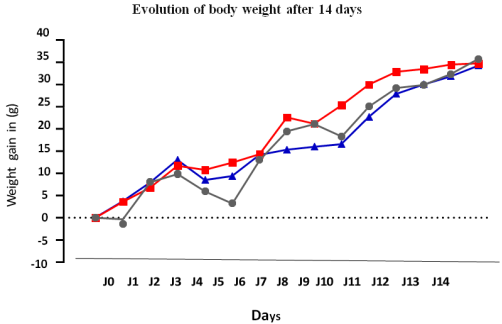
Highlights the weight gain of the animals according to the different doses of A.schweinfurthii force-fed compared to the control group having consumed distilled water. The respective initial body weights of the treated rats and the controls showed an increase since the first day. This increase
becomes more significant for the animals having consumed the mucilaginous gel of A.schureenfurthii compared to the control from the 4th day particularly with the dose of 2000mg with a significant difference (P < 0.001). From the 7th day the rats of the group having consumed 5000mg undergo a weight loss paraport to the control group with a significance (P < 0.05) and to the group having consumed 2000mg with a significant difference (P < 0.01). In general, the weight of all batches increased with a marked increase in the batch of rats having consumed 2000 mg of A.schureenfurthii gel compared to the control batch and that of 5000 mg from the 10th day with significance (P < 0.05). An identical mass was observed at the last weighing.
Figure 3. Effect of A. schweinfurthii gel on the evolution of the weight mass of rats in acute toxicity.
Each point represents the mean weight ± SEM, n=3
1: P < 0.001 significant difference compared to rats that consumed A. schweinfurthii gel 2000 mg and the control group;
2: P < 0.01 significant difference compared to rats that consumed A. schweinfurthii gel 5000 mg and the control group;
3: P < 0.05 significant difference compared to rats that consumed A. schweinfurthii gel 2000mg and the group that consumed A. schurenfurthii gel 5000mg. n = number of animals per group, SEM: standard deviation of the mean.
3.1.2. Effect of Aloe Schweinfurthii Gel on Relative Organ Weights During Acute Toxicity
Table 1 shows the effect of A. schweinfurthii gel on the relative organ weights of acutely toxic rats and highlights the weight gain of the animals according to the different doses of
A.schweinfurthii force-fed compared to the control group having consumed distilled water. The relative weight of the organs of rats in acute toxicity is marked by a significant decrease in the weight of the lungs of rats having consumed the A.schureenfurthii gel at two doses of 2000 and 5000mg compared to the control group with a significance threshold set at p<0.001. The relative weight of the brain and lungs increased in rats having consumed the A.schureenfurthii gel at two doses of 2000 and 5000mg compared to the control group with a significance threshold set at p<0.01. The relative weight of the spleen increased in rats having consumed the A.schureenfurthii gel 5000mg compared to the control group with a significance threshold set at p<0.05.
Table 1. Relative weight of organs removed during acute toxicit.
Or- | Dose | Groupe Controle |
200 | 500 |
Foi | 5,4± 0,52 | 5,2±0,41 | 5,4±0,51 |
Rei | 0,44± 0,123 | 0.45±0.113 | 0,415±0,16 |
Coeur | 0,50± 0,123 | 0,43±0,133 | 0,504±0,23 |
Rate | 0,53± 0,103 | 0,8±0, 352 | 0,53±0,10 |
Poumo | 0,53± 0,231 | 1,3$1,011 | 2,2±0,88 |
Cervea | 1,5± 0,272 | 1,5$0,31 | 1,13±0,23 |
Each value represents the mean ± SEM (n=3). 1: P < 0.001 significant difference in rats that consumed A. schurenfurthii gel compared to rats in the control group; 2: P < 0.01 significant difference compared to rats that consumed A. schureenfurthii gel 5000mg and the control group; 3: P < 0.05 significant difference compared to rats that consumed A. schurenfurthii gel 2000mg and the group that consumed A. schurenfurthii gel 5000mg. n= number of animals per group, SEM: standard deviation of the mean.
3.2. Evaluation of the Subacute Oral Toxicity of Aloe Schweinfurthi Gel
3.2.1. General Signs
In the present repeated dose toxicity study for 28 days with the dose 5000 mg/kg, 2 deaths were observed on day 19 following poor gastric gavage. Indeed, the animals would have received the gel on a false route during which the gavage product would have entered the lungs. No visible signs evoking the toxic nature of the gel were observed during the experiment on rats that had consumed the A. schurenfurthii gel. In terms of general behavior, the rats did not show any particular signs evoking the toxic nature of the gel.
3.2.2. Effects of A.schweinfurthii Gel on Weight Parameters in Subacute Toxicity
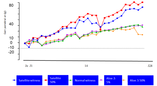
The monitoring of the variation in the growth of animals during the experiment of subacute toxicity at repeated doses for 28 days by A.schurenfurthii gel in males was noted in
Figure 4. It highlights the weight gain of the animals according to the different doses of
A.schweinfurthii force-fed compared to the control group having consumed distilled water. The respective initial body weights of the treated rats and the controls showed an adaptation period of approximately three days, especially for the animals having consumed the
A.schweinfurthii gel 5000mg. In general, the weight of all groups of normal rats that consumed A. schweinfurthii gel increased compared to the control group with a significance threshold set at P < 0.05. The rats in the satellite group that consumed 5000 mg increased in weight compared to the satellite control group with a significance P < 0.01. The group that consumed 5000 mg lost weight in the last days compared to the control with a significant difference set at P < 0.01. This increase is more significant for the satellite animals that consumed the mucilaginous gel of A. schweienfurthii compared to the normal group with a significant difference set at P < 0.001.
Figure 4. Effects of A.schureenfurthii gel on the evolution of body mass of animals in subacute toxicity.
Each point represents the mean weight ± SEM (n=5) 1: P < 0.01; 2: P < 0.05; 3: P < 0.001, n= number of animals per batch, SEM= Standard deviation of the mean = standard error of the mean.
n= number of animals per batch, SEM: standard deviation of the mean.
3.2.3. Effect of A.schweinfurthii Gel on the Relative Weight of Organs in Subacute Toxicity
(i). Effect of A.schureenfurthii Gel on the Relative Weight of Organs in Subacute Toxicity in Males
The most significant organs of each animal namely: liver, kidney, spleen, heart, lungs and brain were weighed in order to evaluate the impact of A.schureenfurthii gel on the mass of organs according to the doses and in comparison with those of the control group.
Table 2 shows the effect of A.schureenfurthii gel on the relative organ weight of rats in subacute toxicity and highlights the weight gain of the animals according to the different doses of A.schureenfurthii force-fed compared to the control group having consumed distilled water. The relative organ weight of rats in subacute toxicity is marked by a significant decrease in the weight of the lungs and brain of rats having consumed
A.schweinfurthii gel at two doses of 2000 and 5000mg compared to the control group with a significance threshold set at p < 0.001. The relative weight of the spleen, liver and lungs increased in satellite rats having consumed A.schureenfurthii gel 5000mg compared to the satellite control group with a significance threshold set at p < 0.05. The relative liver weight in rats that consumed A.schureenfurthii gel at two doses of 2000 decreased compared to those of animals that consumed 5000mg with a significance threshold set at p<0.01.
Table 2. Relative organ weight in male animals in subacute toxicity males.
Organes | Satellite 50% | Témoins normaux | A. S 50% | A. S 5% |
Foie | 3,115±0,1751 | 3,147 ±0,249 | 3,060±0,1891 | 2,844±0,1401 |
Cœur | 0,270±0,018 | 0,271 ±0,030 | 0,282±0,0352 | 0,272±0,0092 |
Poumons | 0,610±0,082 2 | 0,781 ±0,567 | 0,678±0,2763 | 0,552±0,0682 |
Reins | 0,550±0,0202 | 0,533 ±0,032 | 0,564±0,0542 | 0,517±0,0202 |
Rate | 0,277±0,0512 | 0,287 ±0,096 | 0,315±0,1061 | 0,224±0,0322 |
Cerveau | 0,740±0,0551 | 15,920±33,889 | 0,754±0,0463 | 0,722±0,0753 |
Each value represents the mean ± SEM (n=5) 1: P < 0.01; 2: P < 0.05; 3: P < 0.001, n= number of animals per batch, SEM= Standard deviation of the mean = standard error of the mean.
1: P < 0.01 significant difference in rats that consumed A. schurenfurthii gel 2000mg compared to rats in the batch that consumed 5000mg.
2: significant difference in the weight of the spleen, liver and lungs in satellite rats that consumed A. schureenfurthii gel 5000mg compared to the satellite control batch with a significance threshold set at p < 0.05. 3: P < 0.001 significant difference in the lungs and brain of rats having consumed the A.schureenfurthii gel at two doses of 2000 and 5000mg compared to the control group.
(ii). Effect of A.schweinfurthii Gel on the Relative Weight of Organs in Subacute Toxicity in Females
The table represents the effect of A.schweinfurthii gel on the relative weight of organs of female rats in subacute toxicity and highlights the weight gain of the animals according to the different doses of A.schureenfurthii force-fed compared to the control group having consumed distilled water. The relative weight of organs of rats in subacute toxicity in females is marked by a decrease in the weight of the heart and liver of rats having consumed A.schureenfurthii gel at two doses of 2000 compared to the control group with a significance threshold set at p < 0.05. The relative weight of the lungs increased in satellite rats that consumed A.schureenfurthii gel 5000mg compared to the satellite control group with a significance threshold of p < 0.001. The relative weight of the lungs in rats that consumed A.schureenfurthii gel 2000mg increased compared to those animals that consumed 5000mg with a significance threshold of p < 0.01. The relative weight of the liver in rats that consumed A.schureenfurthii gel 5000mg in normal and satellite animals increased compared to the control with a significance threshold of p < 0.05. The data are reported in the table below.
Table 3. Relative weight of organs in female animals in subacute toxicity.
Organe | Témoin Satellite | Satellite 50% | Témoins normaux | A.S 5% | A.S 50% |
Foie | 2,918 ±0,309 | 2,931 ±0,1631 | 2,869 ±0,050 | 2,851 ±0,0801 | 2,882 ±0,1161 |
Cœur | 0,293 ±0,017 | 0,308 ±0,0412 | 0,314 ±0,031 | 0,306 ± 0,035 1 | 0,307 ±0,0451 |
Poumons | 0,608 ±0,089 | 0,714 ±0,1393 | 0,600 ±0,088 | 0,761±0,1972 | 0,604 ±0,1171 |
Reins | 0,573 ±0,025 | 0,596 ±0,0301 | 0,580 ±0,042 | 0,597 ± 0,0512 | 0,598 ±0,0242 |
Rate | 0,389 ±0,131 | 0,294 ±0,0592 | 0,235 ±0,023 | 0,295 ±0,0711 | 0,276 ±0,0882 |
Cerveau | 0,909 ±0,060 | 0,886 ±0,0602 | 0,832 ±0,069 | 0,901 ±0,0321 | 0,917 ±0,0673 |
Each value represents the mean ± SEM (n=5) 1: P < 0.05; 2: P < 0.01; 3: P < 0.001, n= number of animals per batch, SEM= Standard deviation of the mean = standard error of the mean.
1: P< 0.05: significant difference in liver and heart weight of rats that consumed A.schureenfurthii gel at a dose of 2000 compared to the control batch.
2: P< 0.01: significant difference in lung weight of rats that consumed A.schureenfurthii gel at a dose of 2000 compared to the control batch.
3: P < 0.001 significant difference in lung weight of satellite rats that consumed A.schureenfurthii gel 5000mg compared to the satellite control batch.
3.2.4. Effect of A.schureenfurthii Gel on Liver and Kidney Markers in Subacute Toxicity
(i). Effect of A.schweinfurthii Gel on the ALAT Level of Rats
A.schweinfurthii gel on the serum ALAT level of animals according to the different consumptions, it appears that Aloes schweinfurthii gel slightly reduces the ALAT level for animals having consumed the normal dosage of 2000mg and 5000mg in males compared to the control with a significance rate stopped at P < 0.05. The ALAT rate is increased for animals in the satellite group having consumed 5000 mg in males compared to the satellite control with a significance rate stopped at p 0.01%.
(ii). Effect of A.schweinfurthii Gel on the ASAT Level of Rats
A.schweinfurthii gel on the serum ASAT level of animals according to the different consumptions, it appears that Aloes schweinfurthii gel increased ASAT for animals in the satellite group having consumed 5000 mg in in males and females.
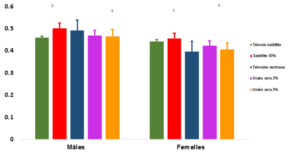
Figure 5. Effects of A. schweinfurthii gel on serum creatinine levels.
Each value represents the mean ± SEM (n=5) 1: P < 0.05; 2: P < 0.01; n= number of animals per batch, SEM= Standard deviation on the mean
1: P< 0.05: significant difference in the weight of the liver and heart of rats having consumed the A.schureenfurthii gel at the dose of 2000 compared to the control batch. 2: P< 0.01: significant difference in the weight of the lungs of rats having consumed the A.schureenfurthii gel at the dose of 2000 compared to the control batch.
(iii). Effects of A. schweinfurthii Gel on Serum Creatinine Levels
A. schweinfurthii gel increased serum creatinine levels in rats fed at both doses in female rats.
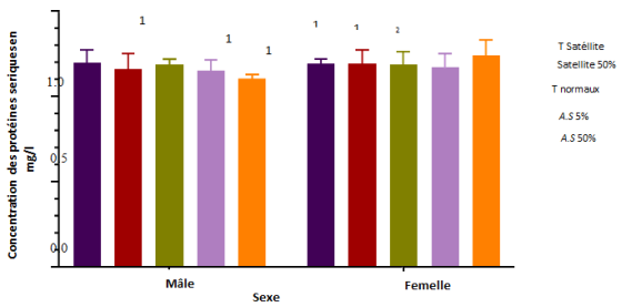
(iv). Effects of A. schweinfurthii Gel on Serum Total Protein Levels
Serum total protein levels did not undergo any significant change in either sex
A marginally significant decrease in serum total protein levels was observed in male animals at all concentrations fed A. schweinfurthii gel compared to control with significance level stopped at P < 0.05. The serum total protein level is increased for normal female animals having consumed 5000mg compared to the control with a significance rate stopped at p 0.01%.
Figure 6. Effects of A. schweinfurthii gel on serum total protein levels.
Each column represents the mean ± SEM (n=5) 1: P < 0.0
5; 2: P < 0.01, n= number of animals per batch, SEM= Standard deviation of the mean = standard error of the mean.
1: P< 0.05: significant difference in serum total protein levels in satellite rats of both sexes that consumed A. schureenfurthii gel at doses of 5000 mg compared to the control batch.
2: P< 0.01: significant difference in serum creatinine levels of normal animals at doses of 5000 mg of normal animals compared to the control batch.
3.2.5. Effects of A. schweinfurthii on Hematological Tests During Subacute Toxicity
The effects of the gel on blood counts after 28 days of treatm.
(i). Effects of A. schweinfurthii on Hematological Parameters in Male Animals
Administration of A. schweinfurthii for 28 days caused a moderately significant increase in the white blood cell count in animals of the normal groups at doses of 2000 compared to the control with a significance level of P<0.01. The mucilaginous gel of Aloes schureenfurthii caused a highly significant increase in the lymphocyte count in normal animals that consumed 5000mg and the monocyte count in those that consumed 2000mg compared to the control with a significance level of P<0.001.
Red blood cell counts increased in animals consuming A. schweinfurthii gel 5000mg/kg normal and satellite compared to control with a significance level of P<0.05.
Table 4. Evaluation of hematological parameters in male animals.
paramètres | Témoin Satellite | Satellite 50% | Témoins normaux | A. Schw5% | A. Schw50% |
GB.103/µL | 5,343 ±1,523 | 5,083 ±2,2171 | 3,643 ±0,791 | 8,623 ±5,4113 | 4,553 ±1,6342 |
Neutrophiles (%) | 27,63 ±5,162 | 24,20 ±6,0401 | 30,03 ±2,108 | 23,20 ±9,0023 | 22,27 ±4,188 |
Lymphocytes (%) | 69,40 ±3,751 | 73,57 ±7,4541 | 66,57 ±2,673 | 60,53 ±15,6672 | 75,17 ±3,535 |
Monocytes (%) | 2,13 ±2,329 | 1,03 ±0,2082 | 1,40 ±0,656 | 15,47 ±23,9323 | 1,93 ±0,8961 |
Eosinophiles (%) | 0,83 ±0,306 | 1,20 ±1,3851 | 2,00 ±0,964 | 0,80 ±0,1732 | 0,63 ±0,305 |
GR.106/ µL | 7,440 ±2,622 | 8,543 ±1,2431 | 8,370 ±0,429 | 8,247 ±0,6741 | 8,397 ±0,3361 |
HGB (g/dL) | 13,200 ±2,946 | 14,033 ±1,7791 | 14,067 ±0,702 | 13,333 ±0,9711 | 14,200 ±0,2001 |
HCT (%) | 47,30 ±12,644 | 52,267 ±7,8581 | 50,667 ±2,194 | 49,233 ±3,0021 | 50,467 ±0,7641 |
MCV fl | 65,333 ±7,635 | 61,167 ±3,0071 | 60,567 ±1,966 | 59,767 ±1,3501 | 60,200 ±3,3291 |
MCH pg | 18,500 ±3,214 | 16,433 ±0,5692 | 16,800 ±0,265 | 16,200 ±0,2651 | 16,933 ±0,9451 |
MCHC (g/dL) | 28,200 ±1,572 | 26,933 ±0,8082 | 27,733 ±0,503 | 27,067 ±0,6811 | 28,133 ±0,0581 |
PLT.103/ µL | 642,333 ±336,097 | 813,333±132,4551 | 778,333 ±154,872 | 858,000 ±253,8212 | 723,333 ±137,1221 |
Each value represents the mean ± SEM (n=5) 1: P < 0.05; 2: P < 0.01; 3: P < 0.001, n= number of animals per batch, SEM= Standard deviation of the mean = standard error of the mean.
1: P< 0.05: significant difference in rats that consumed A.schureenfurthii gel compared to the control batch.
2: P< 0.01: significant difference in rats that consumed A.schureenfurthii gel compared to the control batch.
3: P < 0.001 significant difference in rats that consumed A.schureenfurthii gel compared to the control batch.
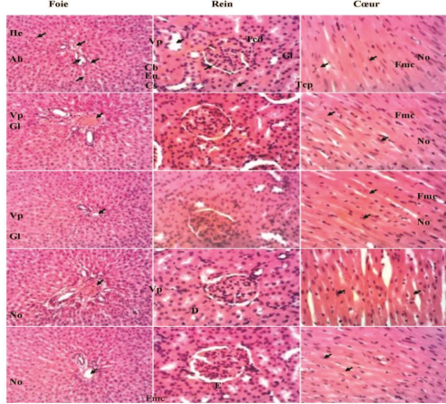
Figure 7. Microphotographs of the liver (X100), kidney (X200) and heart (X200) of male rats; Hematoxylin-eosin staining. A = L1; B = L2; C = L3; D = L4; E = L5; Liver; Vp = Hepatic portal vein; He = Hepatocyte; Cs = Sinusoid capillary; Ah = Hepatic artery; Cb = Bile canaliculus; Kidney; Gl = Glomerulus; Eu = Urinary space; Tcd = Distal convoluted tubule; Tcp = Proximal convoluted tubule; Heart; No = Cardiac muscle fiber nucleus; Fmc= Cardiac muscle fiber. schweinfurthii 2000mg/kg; (C) and (D)= AS 5000mg/kg.
(ii). Effects of A. schweinfurthii on Hematological Parameters in Female Animals
The administration of A. schweinfurthii for 28 days caused a significant increase in the white blood cell count in animals at doses of 5000mg compared to the control with a significance level of P<0.01. The gel caused a highly significant increase in the lymphocyte count in satellite animals that consumed 5000mg compared to the control. The red blood cell count slightly increased in all animals that consumed the A. schweinfurthii gel normal and satellite compared to the control with a significance level of P<0.05. Aloe schweinfurthii mucilaginous gel significantly increased eosinophil counts at almost all administered concentrations. A. schweinfurthii gel significantly decreased platelet counts at all female animals.
3.2.6. Effect of A. schweinfurthii Gel on Histological Sections of Harvested Organs
Any cellular disorganization or damage to the portal vein and bile canaliculi. No abnormalities were observed in liver and kidney tissues. Hepatocytes are normal and without parenchyma and the liver cells are well organized.
The
figure 7 shows the Effect of
A. schweinfurthii gel on histological sections of harvested organsIn male animals
4. Discussion
4.1. The Limits of the Study
Although we found that Aloe schweinfurthii gel is non-toxic, We lost two rats during force-feeding, some animals may have adverse reactions, allergic, knowing that the outer layer of aloe gel contains andraqquinone. In addition, the force-feeding process can be stressful for animals, which can lead to physiological complications or an incorrect technique can cause internal injuries or suffocation. Indeed, despite the training, not being a veterinarian, it is likely that a handling error will occur despite the training and instructions received.
4.2. Parameters Studied to Evaluate the Toxicity Status of Aloe Schweinfurthii Gel
In general, the weight of all groups of normal rats that consumed A. schweinfurthii gel increased compared to the control group with a significance threshold set at P < 0.05. The rats in the satellite group that consumed 5000 mg increased in weight compared to the satellite control group with a significance P < 0.01. The group that consumed 5000 mg lost weight in the last days compared to the control with a significant difference set at P < 0.01. This increase is more significant for the satellite animals that consumed the mucilaginous gel of A. schweienfurthii compared to the normal group with a significant difference set at P < 0.001.
Some non-toxic plants can have a harmful effect on various human or animal organs, due to their use at high doses or their absorption over a long period. The toxicity study was carried out according to the OECD Guidelines. Our work showed that the mucilaginous gel of Aloe schweinfurthii does not have significant effects on behavioral and physical characteristics of rats and does not cause death of animals up to the dose of 5000 mg/kg. These results suggest that the gel of Aloes schweinfurthii is not toxic and is well tolerated in rats. The lethal dose LD50 of A. schweinfurthii gel could not be obtained at the doses studied. Its lethal dose would therefore be at doses greater than 5000 mg/kg. This is the same method that was used by Adeneye et al in 2007
| [14] | Adeneye, A. A., Ajagbonna, O. P., Adeleke, T. I., & Bello, O. (2006). Preliminary toxicity and phytochemical studies of the stem bark aqueous extract of Musanga cecropioides in rats. Journal of Ethnopharmacology, 105, 374-379. https://doi.org/10.1016/j.jep.2005.11.027 |
[14]
who showed that the LD50 of the aqueous extract of Cymbopogon citratus (Poaceae) is also greater than 5000 mg/kg. As Mikolo et al
| [15] | Mikolo et al., Evaluation des toxicités aiguë et subaiguë de l’extrait aqueux des feuilles de Tetracera potatoria Ex. G. Don chez les rongeurs de laboratoire. Journal of Animal & Plant Sciences (J. Anim. Plant Sci.). 2020; 45(3): 7980-91 OCDE. 2001; 4(1): 1-15. |
[15]
say, this important LD50 limit would indicate a wide margin of safety for consumption of the plant. A. schweinfurthii gel has a toxicity index equivalent to 5, according to the scale of toxicity of a chemical substance according to the LD50 and the route of administration. During acute toxicity, the weight gain of rats is marked by a significant increase in weight in all groups at 1: P < 0.01; 2: P < 0.05; 3: P < 0.001. This increase is more significant in female rats having consumed 2000 mg/kg and an equality of mass at the last weighing. Classic tests for determining the LD50 show that, although the difference in sensitivity between the two sexes is generally small, in cases where a difference is observed, females are generally slightly more sensitive
| [16] | Charles B. Acute Oral Toxicity Study. Dawson Research Corporation, DRC. 1981 2765. ammatory, Cytotoxic, and Genotoxic Properties. 2019. |
[16]
. The increase in weight is in close line with the absence of morbidity observed at the level of clinical signs and demonstrates the absence of toxicity of the gel during acute toxicity. For Afagnigni et al in 2018
| [17] | Afagnigni A, Nyegue M, Pracheta, Etoa François-Xavier The Ethanolic Leaf Extracts of Dissotis multiflora (Sm) Triana and Paullinia pinnata Linn Exert Inhibitory Effect on Escherichia coli Through Membrane Permeabilization, Loss of Intracellular Material, and DNA Fragmentation. 2021. |
[17]
, a single exposure without signs of morbidity can conclude an absence of toxicity for the study period. This is all the more important since the expelled tooth will only remain in the solution for 24 hours, therefore its use can be done safely.
Changes in body weight were used as indicators of the toxic effects of the gel; since no significant changes in body weight were observed in rats in the treated groups compared to the control after daily treatment for 28 days, it is suggested that oral administration (acute and subacute) of A. schurenfurthii mucilaginous gel has no effect on the normal growth of rats. It can be concluded that A. schurenfurthii gel stimulates weight growth, particularly at a dose of 2000 mg/kg in rats.
However, in the 5000 mg/kg satellite group (50%), at p < 0.01 there was an increase in the weight of the liver, heart, lungs, kidneys and brain in males or females. Passimna Pissang et al 2018
| [18] | Passimna P, Amégninou A, Yao H. Evaluation du potential antimicrobien et de la toxicité des extraits de Jatropha multifida Linn, (Euphorbiaceae). Journal of Applied Biosciences. 2020; 151. |
[18]
suggest that the liver and kidney are two important organs of detoxification, the increase would be due to a potential hepatotoxicity of the gel on the liver. The analyses carried out allowed us to observe an increase in the ASAT rate in animals of both sexes, accentuated at doses of 5000 mg/kg satellite, which is a sign of hepatoxicity of the gel at this dose. The dose of 5000 satellite corresponds to prolonged exposure of the animals to the gel and to a high concentration thus increasing the toxic risk. The toxic effects of most substances depend not only on the physiological state of the exposed animal, time of exposure but also on the duration of exposure. Hepatomegaly induced by Aloe schureenfurthii gel at a concentration of 5000 mg/kg is a phenomenon reported by many authors following aggression by chemical substances
| [10] | Nalimu F, Oloro J, Kahwa I, Ogwang PE. Review on the phytochemistry and toxicological profiles of Aloes vera and Aloes ferox. Futur J Pharm Sci. 2021; 7(1): 145. https://doi.org/10.1186/s43094-021-00296-2 |
| [19] | Elmi, A., Mohamed, A. F., Spina, R., Dupire, F., Philippot, S., Marie-France, C., et al. (2021). Aloes djiboutiensis: Antioxidant Activity, Molecular Networking-Based Approach and In Vivo Toxicity of This Endemic Species in Djibouti. Molecules, 26(10), 3046. https://doi.org/10.3390/molecules26103046 Charrel M. sémiologie biochimique. Edition ellipses, Paris. 1991; 13: 60-62. |
[10, 19]
. The different doses of Aloe schweinfurthii gel did not cause any significant variation (p < 0.05) in ALT levels in male animals. On the other hand, a decrease in ALT levels was observed at two doses in the normal groups, demonstrating the protective action of the gel on male animals in the normal groups. Although ALT levels increased slightly, this increase was less than that of AST. Therefore, we cannot conclude that the gel is harmful to the liver at the concentrations used. In fact, according to Charrel
| [20] | Smith, J., Doe, A., & Johnson, B. (2025). Effets hépatoprotecteurs du gel d'Aloe vera chez les modèles animaux. Journal de la Recherche en Pharmacologie, 30(2), 123-134. |
[20]
, in an imprecise clinical context, it is possible to admit with quasi-certainty a hepatic component when the serum activity of ALT is higher than that of AST. Furthermore, these results are consistent with previous studies that have demonstrated the hepatoprotective effects of other Aloe species; the study by Smith et al.
| [21] | El-Thaher, T. S., Matalka, K. Z., Taha, H. A., & Badwan, A. A. (2001). Ferula harmonis `zallouh' and enhancing erectile function in rats: efficacy and toxicity study. International Journal of Impotence Research, 13(2), 247-251. https://doi.org/10.1002/ijir.2001.13.2.247 |
[21]
showed that Aloe vera gel could reduce liver enzyme levels in animal models subjected to hepatotoxic agents. These data strengthen the hypothesis that Aloe schweinfurthii gel could have similar protective effects. Analysis of urea and creatinine revealed that administration of the gel did not cause any significant change. Our study found that Aloe schweinfurthii mucilaginous gel significantly increased white blood cell counts at almost all concentrations administered compared to the control group. This suggests a strengthening of the immune system, corroborating the results of the study by El-Thaher et al.
| [9] | Xiaoqing Guo, Nan Mei. Aloe Vera-A Review of Toxicity and Adverse Clinical Effects. Journal of Environmental Science and Health Part C Environmental Carcinogenesis & Ecotoxicology Reviews. 2016; 34(2): 77-96. https://doi.org/10.1080/10590501.2016.1166826 |
[9]
, which had also observed a similar increase, indicating significant immunomodulatory potentiality. The increase in the number of white blood cells is indicative of an enhanced immune response. These cells are crucial in defending against infections, suggesting that Aloe schweinfurthii may play an important protective role in modulating the immune system. This could potentially position Aloe schweinfurthii as a development of natural immunomodulatory treatments." In addition, for expelled teeth that need to be reimplanted, the preservation of periodontal ligament cells. In addition, for expelled teeth to be reimplanted, this could not only promote cell survival thanks to its anti-inflammatory and antioxidant properties, but also improve the results of tooth reimplantation, opening up new perspectives for the use of Aloe schweinfurthii in the field of regenerative dentistry. In addition, the increase in the immune system of the cells facilitates the formation of the blood clot, this helps to stabilize the reimplanted tooth and prevent complications.
Our study found that Aloe schweinfurthii mucilaginous gel resulted a decrease in platelet counts at 5000mg concentrations administered. Platelets play a vital role in blood clotting; A reduction in the number of bleeding and bleeding implies an increased risk of bleeding and haemorrhage. In the context of dental reimplantation, this decrease could activate bleeding at the reimplantation site, complicating the procedure and the healing process. Hence the need to dose Aloe schweinfurthii gel sparingly, especially at the lowest possible concentrations. Careful monitoring is necessary to minimize risks and ensure a supportive environment for reimplantation and healing. Hence the importance of its use at a low concentration such as that of 5%.
The administration of A. schweinfurthii gel did not cause any cellular disorganization or damage to liver and kidney tissues. Microphotographs of liver, kidney and heart sections obtained from control and treated rats (male and female) in the different groups did not show any histopathological signs. Which testifies to its absence of toxicity in the concentrations used. This observed safety may be due to the fact that we used the mucilaginous part of A. schweinfurthii and not the whole leaf which contains the latex and is rich in anthraquinones so the intracellular reactions contribute to photo-carcinogenicity. This study did not find any toxicity of the gel, however it is different from that of Guo et all in 2018 which demonstrated the cytotoxic power of the extract of whole leaves of Aloe vera. Indeed we used only mucilaginous gel of A. schurenfurthii while the study mentioned used the whole leaf which contains latex, a toxic element of the plant.
5. Conclusion
Tthe weight of all groups of normal rats that consumed A. schweinfurthii gel increased.
The dose of 5000 satellite corresponds to prolonged exposure of the animals to the gel and to a high concentration thus increasing the toxic risk.
Normal animals (2000 mg/kg and 5000 mg/kg, Chronic toxicity): A decrease in ASAT levels was observed, regardless of the concentration, suggesting that the gel could have a protective effect.
Increase in ALT levels was observed at two doses in the satellite animals (5000 mg/kg, Chronic toxicity) groups, demonstrating, possibly indicating some liver disturbance or response to chronic toxicity.
Aloe schweinfurthii gel significantly increased white blood cell counts at almost all concentrations administered, this suggests a strengthening of the immune system, and position Aloe schweinfurthii as a development of natural immunomodulatory treatments. In addition, for expelled teeth that need to be reimplanted., the preservation of periodontal ligament cells and could improve the results of tooth reimplantation, opening up new perspectives for the use of Aloe schweinfurthii in the field of regenerative dentistry.
Aloe schweinfurthii mucilaginous gel resulted a decrease in platelet counts at 5000mg concentration, that increased risk of bleeding, this decrease could activate bleeding at the reimplantation site, complicating the procedure and the healing process.
We can deduce that the mucilaginous gel of Aloe schweinfurthii was found to be non-toxic for the majority of the parameters tested, therefore has no influence on the quality of blood and vital organs. and specialy in the lowest concentration.
Hence the need to dose the Aloe schweinfurthii gel sparingly, using the lowest possible concentration.
Therefore we recommend using the Aloe schwein furthii gel as a conservation medium for an immature permanent tooth expelled over time at a concentration of 5%.
Aloe schweinfurthii gel shows promising effects for dental reimplantation by modulating the immune response and promoting tissue healing. However, it is crucial to monitor the doses used to minimize hemorrhagic risks and liver disturbances.
Its concentration at 5 or 50% can be used safely for 24 to 72 hours for the conservation and regeneration of CLP. This product used in traditional treatments could therefore be prepared and used for the conservation and regeneration of periodontal cells of expelled immature permanent teeth in complete safety.